- Home
- Fachgebiete
- Ärzteteam
- Patienten
- Die Klinik Gut
- Jobs & Karriere
- News


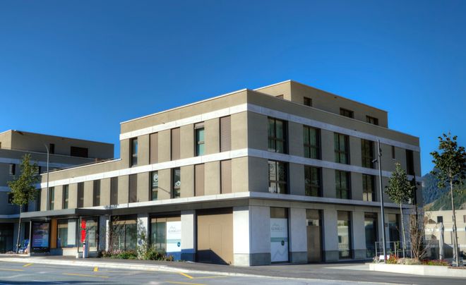
Klinik Gut St. Moritz
Plazza Paracelsus 2a
CH-7500 St. Moritz
+41 81 836 34 34
stmoritz@klinik-gut.ch
24h-Notfalldienst
Bushaltestelle: 'St. Moritz Bad, Via San Gian' oder 'St. Moritz Bad, Hallenbad'
(Wir sind umgezogen. Die Bushaltestelle 'Klinik Gut' an der Via Serlas bedient unseren früheren Standort in St. Moritz-Dorf.)
Praxis Klinik Gut Chur
Masanserstrasse 237
CH-7000 Chur
+41 81 258 4400
chur@klinik-gut.ch
offen 8–12 und 14–17 Uhr (Mo–Fr)
Besucherparkplätze entlang des Gebäudes (Seite Rheingässli) oder in der Tiefgarage (Zufahrt über das Rheingässli)
Route Planen
Klinik Gut Fläsch
Steigstrasse 12
CH-7306 Fläsch
+41 81 595 55 55
flaesch@klinik-gut.ch
Besucherparkplätze vorhanden (Tiefgarage und vor dem Haus)
Route Planen
Praxis Klinik Gut Zürich
Airport Medical Center
CH-8060 Zürich-Flughafen
+41 43 816 60 00
amc@klinik-gut.ch
Öffnungszeiten:
täglich von 7.30 Uhr bis 20.30 Uhr (Ärzte der Klinik Gut sind jeden Freitag vor Ort. Wir bitten Sie, telefonisch einen Termin zu vereinbaren.)

Praxis Klinik Gut Ascona
Via Baraggie 3
CH-6612 Ascona
+41 91 780 05 27
ascona@klinik-gut.ch
Öffnungszeiten:
8.30 Uhr bis 17.30 Uhr, jeweils einen Montag im Monat
Praxis Klinik Gut Buchs
Fichtenweg 10
CH-9470 Buchs (SG)
+41 81 599 92 00
buchs@klinik-gut.ch
Öffnungszeiten:
Montag, Dienstag und Donnerstag 08.00 bis 12.00 und 14.00 bis 17.00 Uhr
Mittwoch und Freitag 08.00 bis 12.00 Uhr
Wir nehmen den Schutz Ihrer Personendaten sehr ernst. Die Beachtung der Privatsphäre und der vertrauensvolle Umgang mit ihren Personendaten ist uns ein wichtiges Anliegen. Das Einhalten der gesetzlichen Bestimmungen zum Datenschutz ist für uns selbstverständlich. Nachfolgend geben wir Ihnen einen Überblick darüber, wie wir den Schutz Ihrer Personendaten gewährleisten und welche Ihrer Daten wir zu welchem Zweck bearbeiten.
Für die in dieser Datenschutzerklärung beschriebenen Datenbearbeitungen ist die Klinik Gut verantwortlich. Wenn Sie dazu Fragen haben oder Ihre datenschutzrechtlichen Betroffenenrechte wahrnehmen möchten, können Sie uns dies an folgende Adresse mitteilen:
Klinik Gut AG
Plazza Paracelsus 2a
7500 St. Moritz
Schweiz
+41 81 836 34 34
info@klinik-gut.ch
Folgende Arten von Personendaten bearbeiten wir:
Wir bearbeiten Ihre Personendaten zu folgenden Zwecken:
Die Rechtsgrundlage für die Bearbeitung Ihrer Personendaten hängt im Einzelfall vom jeweiligen Zweck der Datenbearbeitung ab. In Frage kommen namentlich:
Wir bearbeiten und speichern wir Ihre Personendaten nur in dem Umfang und so lange, wie es für die Erfüllung unserer vertraglichen und gesetzlichen Pflichten oder sonst für die mit der Bearbeitung verfolgten Zwecke erforderlich ist, d.h. also zum Beispiel für die Dauer des gesamten Behandlungsvertrages sowie darüber hinaus gemäss den gesetzlichen Aufbewahrungsfristen (insbesondere aus der öffentlich-rechtlichen Gesundheits-, der Kranken- und Unfallversicherungsgesetzgebung) und Dokumentationspflichten. Sobald Ihre Personendaten für die oben genannten Zwecke nicht mehr erforderlich sind oder eine vorgeschriebene Aufbewahrungsfrist abläuft, werden Ihre Personendaten grundsätzlich und soweit möglich gelöscht oder gesperrt.
Die Sicherheit Ihrer Personendaten ist uns wichtig. Wir treffen angemessene sowie geeignete technische und organisatorische Massnahmen, um die Sicherheit Ihrer Personendaten zu wahren und diese gegen unberechtigte oder unrechtmässige Bearbeitung und/oder gegen unbeabsichtigten Verlust, Veränderung, Bekanntmachung oder Zugriff zu schützen. Hierzu gehört unter anderem die Verwendung von anerkannten Verschlüsselungsverfahren (z.B. SSL-Verschlüsselung). Der Zugriff auf Ihre Personendaten wird ausschliesslich denjenigen Mitarbeitern, Dienstleistern oder Partnern von uns gewährt, die diesen Zugriff zur Erfüllung eines Geschäftszwecks oder zur Ausübung ihrer Pflichten benötigen.
Auch den eigenen, unternehmensinternen Datenschutz nehmen wir ernst. Unsere Mitarbeitenden und die von uns beauftragten Dienstleistungsunternehmen sind zur Verschwiegenheit und zur Einhaltung der datenschutzrechtlichen Bestimmungen verpflichtet. Überdies wird diesen der Zugriff auf Ihre Personendaten nur soweit notwendig gewährt.
Wir behandeln Ihre Personendaten grundsätzlich vertraulich und geben sie nur weiter, wenn Sie dem ausdrücklich zugestimmt haben, wir rechtlich dazu verpflichtet oder berechtigt sind oder dies zur Durchsetzung unserer Rechte, insbesondere zur Durchsetzung von Ansprüchen aus dem Vertragsverhältnis, erforderlich ist. Dabei werden die rechtlichen Vorschriften zur Weitergabe von Personendaten an Dritte selbstverständlich eingehalten.
Zur Erbringung unserer Leistungen, zur Einhaltung vertraglicher oder gesetzlicher Vorschriften oder für die weiteren in dieser Datenschutzerklärung genannten Zwecke kann es notwendig sein, dass wir Ihre Personendaten an folgende Kategorien von Empfängern bekannt geben:
Sofern wir Dritte beiziehen, um unsere Leistungen bereitzustellen, ergreifen wir geeignete rechtliche Vorkehrungen sowie entsprechende technische und organisatorische Massnahmen, um für den Schutz Ihrer Personendaten gemäss den einschlägigen gesetzlichen Vorschriften zu sorgen.
Die Bekanntgabe erfolgt in der Regel innerhalb der Schweiz oder an Empfänger in Mitgliedsstaaten der EU bzw. des EWR oder in anderen Staaten mit angemessener Datenschutzgesetzgebung. Eine allfällige Bekanntgabe an Empfänger in weiteren Staaten nehmen wir gestützt auf anerkannte Garantien (insbes. vertragliche Vereinbarungen) oder entsprechend Ihrer Einwilligung im Einzelfall vor.
Sie haben das Recht, von uns Auskunft darüber zu verlangen, ob und wenn ja, welche Personendaten wir von Ihnen bearbeiten.
Sie haben das Recht, die Berichtigung Ihrer unrichtigen Personendaten und gegebenenfalls die Vervollständigung unvollständiger Personendaten in unseren Systemen zu verlangen.
Sie haben das Recht zu verlangen, dass Ihre Personendaten gelöscht werden, zum Beispiel wenn die Daten für die verfolgten Zwecke nicht mehr benötigt werden. Falls wir jedoch verpflichtet oder berechtigt sind, Ihre Personendaten aufgrund gesetzlicher oder vertraglicher Pflichten dennoch zu behalten, können wir in diesen Fällen Ihre Personendaten daher nur soweit erforderlich einschränken bzw. sperren.
Sie haben das Recht, von uns die Einschränkung der Bearbeitung Ihrer Personendaten zu verlangen.
Sie haben gegebenenfalls das Recht, Ihre Personendaten, die wir aufgrund Ihrer Einwilligung oder zur Erfüllung eines Vertrags automatisiert bearbeiten, in einem strukturierten, gängigen und maschinenlesbaren Format zu erhalten bzw. die Übermittlung dieser Daten an einen Dritten zu verlangen. Sofern Sie die direkte Übertragung der Personendaten an einen anderen Verantwortlichen verlangen, erfolgt dies nur, soweit dies technisch machbar ist.
Sie haben das Recht, der Bearbeitung Ihrer Personendaten jederzeit gemäss den gesetzlichen Vorgaben zu widersprechen. Insbesondere haben Sie das Recht auf Widerspruch gegen die Bearbeitung Ihrer Personendaten zum Zwecke der Direktwerbung.
Sie haben das Recht, Ihre Einwilligung zur Bearbeitung Ihrer Personendaten, grundsätzlich mit Auswirkung für die Zukunft, jederzeit zu widerrufen.
Sie haben zudem das Recht auf Beschwerde bei einer zuständigen Aufsichtsbehörde, wenn Sie der Ansicht sind, dass die Bearbeitung Ihrer Personendaten gegen datenschutzrechtliche Bestimmungen verstösst.
Bitte beachten Sie, dass für diese Rechte Ausnahmen gelten. Insbesondere müssen wir Ihre Personendaten gegebenenfalls weiterbearbeiten und speichern, um einen Vertrag mit Ihnen zu erfüllen, eigene schutzwürdige Interessen wie etwa die Geltendmachung, Ausübung oder Verteidigung von Rechtsansprüchen zu wahren, oder aber um gesetzliche Verpflichtungen einzuhalten. Soweit rechtlich zulässig, können wir daher Ihre datenschutzbezogenen Begehren, z.B. Auskunfts- und Löschungsbegehren, auch ablehnen oder diesen nur eingeschränkt entsprechen.
Beim Besuch unserer Website speichern unsere Server temporär jeden Zugriff in einer Protokolldatei. Folgende Nutzer- und Gerätedaten sowie Personendaten werden dabei ohne Ihr Zutun erfasst und von unserem Hoster (ISP - Internet Service Provider) gespeichert:
Die Erhebung und Bearbeitung dieser Daten erfolgt ausschliesslich zum Zweck, die Nutzung unserer Website zu ermöglichen (Verbindungsaufbau), die Systemsicherheit und -stabilität dauerhaft zu gewährleisten, zur Optimierung unseres Internetangebots sowie zu internen statistischen Zwecken. Hier in besteht unser berechtigtes Interesse an der Datenbearbeitung. Eine Zusammenführung dieser Daten mit anderen Datenquellen wird nicht vorgenommen. Zusätzlich sind diese Daten nicht auf einzelne Personen zurückführbar. Wir behalten uns vor, diese Daten zu prüfen, wenn uns konkrete Anhaltspunkte für eine rechtswidrige Nutzung bekannt werden.
Sie haben die Möglichkeit, mit uns (z.B. per Kontaktformular, E-Mail, Telefon oder via sozialer Medien) in Kontakt zu treten. In diesem Fall werden die von Ihnen gemachten Angaben zum Zwecke der Bearbeitung Ihrer Anfrage und deren Abwicklung bearbeitet. Welche Daten im Falle eines Kontaktformulars erhoben werden, ist auf dem jeweiligen Kontaktformular ersichtlich. Die mit (*) gekennzeichneten Angaben sind Pflichtfelder. Alle weiteren Angaben kann die anfragende Person freiwillig mitteilen.
Sie können dieser Datenbearbeitung jederzeit durch eine E-Mail an info@klinik-gut.ch widersprechen. In einem solchen Fall wird Ihre Anfrage nicht weiter bearbeitet.
Ihre Personendaten werden gelöscht, sobald die von Ihnen gestellte Anfrage erledigt ist. Dies ist dann der Fall, wenn sich aus den Umständen entnehmen lässt, dass der betroffene Sachverhalt abschliessend geklärt ist und der Löschung keine gesetzlichen Aufbewahrungspflichten entgegenstehen.
Wenn Sie sich auf eine Stelle bei uns bewerben, bearbeiten wir diejenigen Personendaten, die wir im Rahmen des Bewerbungsverfahrens von Ihnen erhalten. Dazu gehören neben Ihren Angaben zur Person, Ausbildung, Arbeitserfahrung und Fähigkeiten, die üblichen Korrespondenzdaten wie Postanschrift, E-Mail-Adresse und Telefonnummer. Zudem werden alle von Ihnen im Zusammenhang mit der Bewerbung eingereichten Unterlagen, wie Motivationsschreiben, Lebenslauf und die Zeugnisse bearbeitet. Daneben können uns Bewerber freiwillig zusätzliche Informationen zukommen lassen. Diese Daten werden ausschliesslich im Rahmen Ihrer Bewerbung gespeichert, ausgewertet, bearbeitet oder intern weitergeleitet. Ferner können sie für statistische Zwecke (z.B. Reporting) bearbeitet werden. In diesem Fall sind keine Rückschlüsse auf einzelne Personen möglich.
Die Bearbeitung kann auch auf anderem elektronischem Wege erfolgen. Dies ist insbesondere dann der Fall, wenn Sie entsprechende Bewerbungsunterlagen auf dem elektronischen Wege, beispiels- weise per E-Mail, an uns übermitteln.
Die Bearbeitung Ihrer Bewerberdaten erfolgt zur Erfüllung unserer (vor)vertraglichen Verpflichtungen im Rahmen des Bewerbungsverfahrens.
Sie können dieser Datenbearbeitung jederzeit widersprechen und Ihre Bewerbung zurückziehen. Senden Sie Ihren Widerspruch bitte an die in der Stellenanzeige als Ansprechpartner benannte Person oder an die E-Mail-Adresse: info@klinik-gut.ch.
Schliessen wir einen Arbeitsvertrag mit Ihnen, werden die übermittelten Daten zum Zwecke der Abwicklung des Arbeitsverhältnisses unter Beachtung der gesetzlichen Vorschriften gespeichert.
Endet das Bewerbungsverfahren ohne Anstellung, werden Ihre Personendaten gelöscht, sofern Sie uns keine Einwilligung gegeben haben, Ihre Angaben für weitere Bewerbungsverfahren bei uns zu verwenden und gegebenenfalls Sie wieder zu kontaktieren. Sie haben die Möglichkeit, diese Einwilligung nachträglich jederzeit zu widerrufen. Ihren Widerruf können Sie an die E-Mail-Adresse info@klinik-gut.ch oder an die bei der Stellenausschreibung angegebene E-Mail-Adresse senden.
Wir bearbeiten Personendaten zudem im jeweils erforderlichen Umfang zur Erfüllung unserer vertraglichen und vorvertraglichen Verpflichtungen sowie für die Durchführung weiterer von Ihnen angefragten Dienstleistungen, wie dies in dieser Datenschutzerklärung beschrieben ist. Die hierbei bearbeiteten Personendaten, die Art, der Umfang und der Zweck der jeweils erforderlichen Bearbeitung, bestimmen sich daher nach dem jeweils mit Ihnen vereinbarten Vertrag oder von Ihnen angefragten Dienste.
Speichern wir Ihre Personendaten aufgrund einer Vertragsbeziehung, bleiben diese Daten mindestens so lange gespeichert, wie die Vertragsbeziehung besteht und längstens so lange Verjährungsfristen für mögliche Ansprüche von uns laufen oder gesetzliche oder vertragliche Aufbewahrungspflichten bestehen.
In bestimmten Fällen setzen wir sogenannte Cookies ein. Cookies sind kleine Textdateien, die mit Hilfe des Browsers auf Ihrem Rechner abgelegt und gespeichert werden. Diese richten auf Ihrem Rechner keinen Schaden an. Sie können keine Programme ausführen und keine Viren übertragen. Cookies dienen dazu, unser Angebot nutzerfreundlicher, effektiver und sicherer zu machen.
Die meisten der von uns verwendeten Cookies sind sogenannte Session-Cookies. Diese werden automatisch gelöscht, wenn Sie sich ausloggen oder den Browser schliessen. Andere Cookies bleiben über den jeweiligen Nutzungsvorgang hinaus auf Ihrem Computer gespeichert und ermöglichen uns oder unseren Partnerunternehmen (Cookies von Drittanbietern), Ihren Browser beim nächsten Besuch wiederzuerkennen und allfällige Einstellungen von Ihnen (z.B. Sprache, Schriftgrösse und andere Anzeigepräferenzen) über einen bestimmten Zeitraum zu «merken». Soweit andere Cookies (z.B. Cookies zur Analyse Ihres Surfverhaltens) gespeichert werden, werden diese in dieser Datenschutzerklärung gesondert behandelt.
Die meisten Internetbrowser sind regelmässig so eingestellt, dass sie Cookies akzeptieren. Wollen Sie dies nicht, können Sie Ihren Browser so einrichten, dass er Sie über das Setzen von Cookies informiert und Sie die Annahme von Cookies für bestimmte Fälle nur im Einzelfall erlauben oder generell ausschliessen. Sie können auch das automatische Löschen der Cookies beim Schliessen des Browsers aktivieren. Ausserdem können Sie bereits gesetzte Cookies jederzeit über einen Internetbrowser oder andere Softwareprogramme löschen.
Die Vorgehensweise beim Kontrollieren und Löschen von Cookies ist von dem von Ihnen verwendeten Internetbrowser abhängig. Informationen dazu finden Sie im Hilfe-Menü Ihres Browsers. Bitte beachten Sie, dass einzelne Funktionen unserer Website möglicherweise nicht funktionieren, wenn Sie die Verwendung von Cookies deaktivieren.
Auf unserer Website verwenden wir sogenannte Tracking-Tools. Mit diesen Tracking-Tools wird Ihr Surfverhalten auf unserer Website beobachtet. Diese Beobachtung erfolgt zum Zwecke der bedarfsgerechten Gestaltung und fortlaufenden Optimierung unserer Website. In diesem Zusammenhang werden pseudonymisierte Nutzungsprofile erstellt und kleine Textdateien, die auf Ihrem Computer gespeichert sind («Cookies»), verwendet.
Hierzu können Dritt-Unternehmer ebenfalls permanente Cookies, Pixel-Tags oder ähnliche Technologien einsetzen. Der Dritt-Unternehmer erhält von uns keine Personendaten, kann jedoch Ihre Nutzung unserer Website verfolgen, diese Angaben mit Daten anderer Websites, die Sie besucht haben und ebenfalls vom Dritt-Unternehmer verfolgt werden, kombinieren, und diese Erkenntnisse für eigene Zwecke (z.B. Steuerung von Werbung) verwenden. Die Bearbeitung Ihrer Personendaten durch den Dritt-Unternehmer erfolgt dann in Verantwortung des Dienstleisters nach dessen Datenschutzbestimmungen.
Folgende Trackingtools kommen zum Einsatz:
Google Analytics ist ein Dienst von Google LLC, 1600 Amphitheatre Parkway, Mountain View, CA 94043, USA. Bei Nutzern, die ihren gewöhnlichen Aufenthalt im Europäischen Wirtschaftsraum oder der Schweiz haben, ist laut Google die Google Ireland Limited, Gordon House, Barrow Street, Dublin 4, Irland, der für Ihre Daten zuständige Verantwortliche.
Google Analytics verwendet Cookies und ähnliche Technologien, die auf Ihrem Rechner gespeichert werden und die Analyse Ihrer Nutzung dieser Website erlauben. Die durch diese Cookies erzeugten Informationen über Ihre Benutzung dieser Website (einschliesslich Ihrer IP-Adresse) werden an einen Server von Google, möglicherweise in den USA oder anderen Drittstaaten, übertragen und dort gespeichert. Google wird diese Informationen benutzen, um Ihre Nutzung der Website für uns auszuwerten, um Reports über die Websiteaktivitäten für uns zusammenzustellen und um weitere mit der Website- und der Internetnutzung verbundenen Dienstleistungen zu erbringen. Auch wird Google diese Informationen gegebenenfalls an Dritte übertragen, sofern dies gesetzlich vorgeschrieben oder soweit Dritte diese Daten im Auftrag von Google bearbeiten. Google wird, nach eigenen Angaben, in keinem Fall Ihre IP-Adresse mit anderen Daten von Google in Verbindung bringen.
Wir möchten Sie darauf hinweisen, dass unsere Website zur Verbesserung des Datenschutzes Google Analytics nur mit aktivierter IP-Anonymisierung verwendet. IP-Adressen werden daher nur gekürzt weiterbearbeitet. Damit ist eine Personenbeziehbarkeit bei der Analyse der Nutzung unserer Website ausgeschlossen.
Sie können die Speicherung der Cookies durch eine entsprechende Einstellung Ihrer Browser- Software verhindern; wir weisen Sie jedoch darauf hin, dass Sie in diesem Fall gegebenenfalls nicht sämtliche Funktionen dieser Website vollumfänglich nutzen können. Sie können darüber hinaus die Erhebung der Daten sowie die Bearbeitung der Daten durch Google verhindern, indem Sie das unter dem folgenden Link verfügbare Browser Add-On herunterladen und installieren https://tools.google.com/dlpage/gaoptout. Sie können Google Analytics für Display-Werbung deaktivieren und die Anzeigen im Google Display-Netzwerk anpassen, indem Sie die Anzeigeneinstellungen aufrufen: https://adssettings.google.de.
Weitere Informationen zu den Nutzungsbedingungen und zum Datenschutz von Google Analytics finden Sie unter https://marketingplatform.google.com/about/analytics/terms/de/ und https://policies.google.com/privacy.
Wir unterhalten neben dieser Website auch Präsenzen in unterschiedlichen sozialen Medien, welche Sie über entsprechende Schaltflächen auf unserer Website erreichen können. Soweit Sie eine solche Onlinepräsenz besuchen, werden ggf. personenbezogene Daten an den Anbieter des sozialen Netzwerks übermittelt. Wir weisen Sie darauf hin, dass hierbei Nutzerdaten auch an einen Server in einem Drittland übermittelt werden und damit ausserhalb der Schweiz bzw. der EU/ des EWR bearbeitet werden können.
Ferner werden die Daten der Nutzer innerhalb sozialer Netzwerke im Regelfall für Marktforschungs- und Werbezwecke bearbeitet. Zu diesen Zwecken werden im Regelfall Cookies auf den Rechnern der Nutzer gespeichert, in denen das Nutzungsverhalten und die Interessen der Nutzer gespeichert werden. Ferner können in den Nutzungsprofilen auch Daten unabhängig der von den Nutzern verwendeten Geräte gespeichert werden (insbesondere, wenn die Nutzer Mitglieder der jeweiligen Plattformen sind und bei diesen eingeloggt sind).
Für eine detaillierte Darstellung der jeweiligen Verarbeitungsformen und der Widerspruchsmöglichkeiten (Opt-out) verweisen wir auf die nachfolgend verlinkten Datenschutzerklärungen und Angaben der Betreiber der jeweiligen Netzwerke:
Auch im Fall von Auskunftsanfragen und der Geltendmachung von Betroffenenrechten weisen wir darauf hin, dass diese am effektivsten bei den Anbietern geltend gemacht werden können. Nur die Anbieter haben jeweils Zugriff auf die Daten der Nutzer und können direkt entsprechende Massnahmen ergreifen und Auskünfte geben. Sollten Sie dennoch Hilfe benötigen, dann können Sie sich an uns wenden.
Unsere Website kann Links zu anderen Websites enthalten, die nicht von uns betrieben werden und auf die sich diese Datenschutzerklärung nicht erstreckt. Wir haben keinen Einfluss darauf, dass deren Betreiber die Datenschutzbestimmungen einhalten und übernehmen daher auch keine Verantwortung für Richtigkeit, Aktualität und Vollständigkeit der dort bereitgestellten Informationen.
Wir behalten uns ausdrücklich das Recht vor, diese Datenschutzerklärung jederzeit zu ergänzen oder zu ändern. Es gilt die jeweils auf unserer Website veröffentlichte Fassung.
Diese Seite wurde letztmals geändert am 23.06.2021. Wenn Sie Fragen oder Kommentare zu unseren rechtlichen Hinweisen oder zum Datenschutz haben, nehmen Sie bitte unter info@klinik-gut.ch mit uns Kontakt auf.
Wir setzen Cookies (eigene und von Drittanbietern) ein, um Ihnen die Nutzung unserer Webseiten zu erleichtern. Mit der weiteren Nutzung unserer Webseiten sind Sie mit dem Einsatz der Cookies einverstanden. Weitere Informationen zu Cookies entnehmen Sie bitte unserer Datenschutzerklärung.